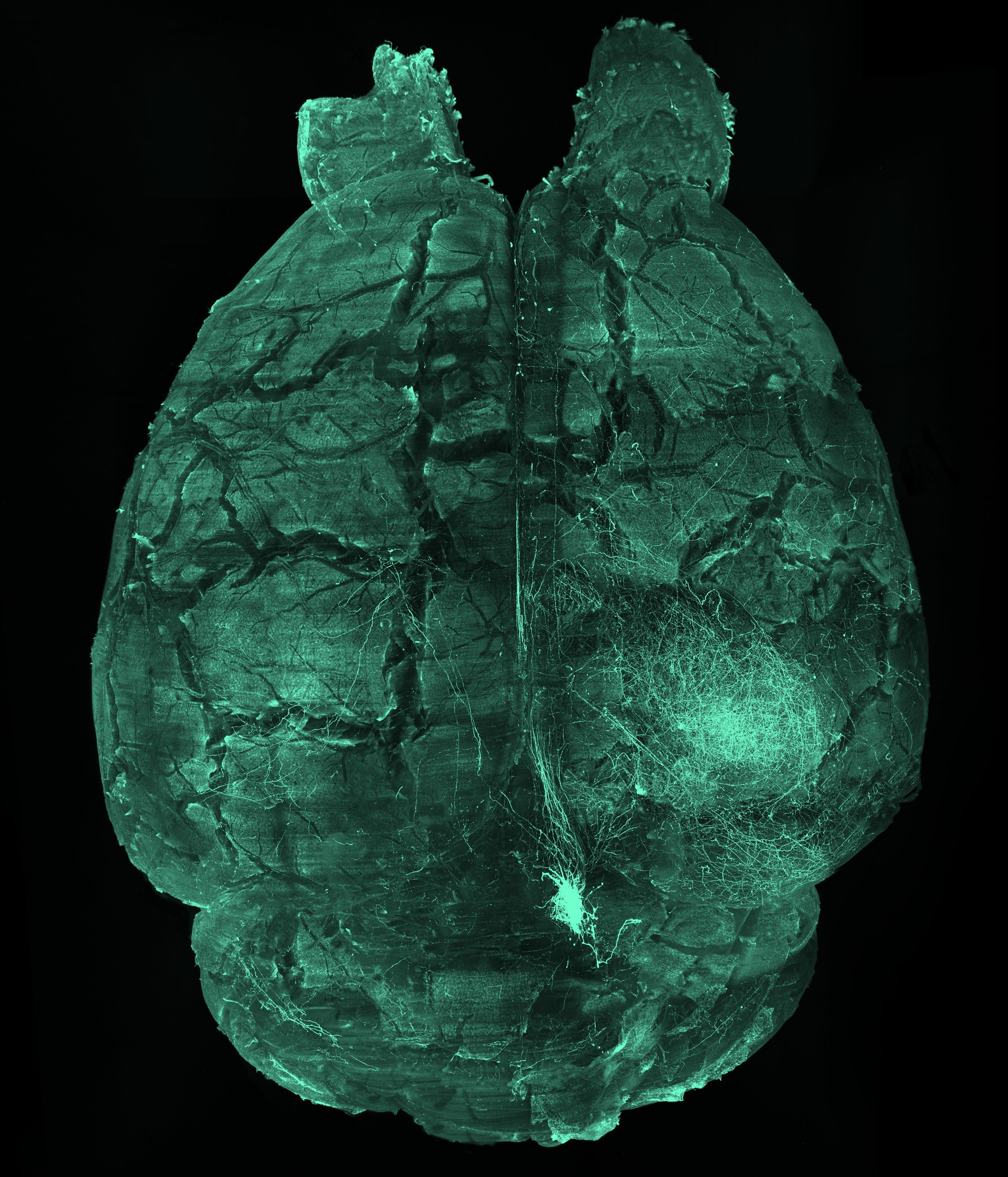
Tract-selective vulnerability in neurodegeneration
We will investigate why particular neuromodulatory projections, including locus coeruleus pathways, show early dysfunction while neighboring circuits remain comparatively resilient.
Future independent research program
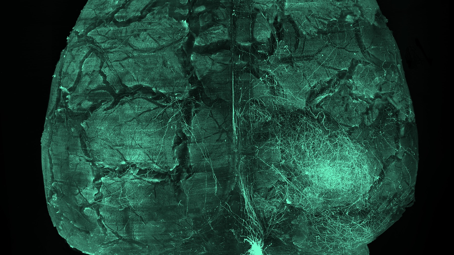
We aim to understand why some neural circuits fail early in neurodegenerative disease while others remain resilient by linking projection-resolved 3D anatomy, glial states, and spatial molecular programs across scales.
The planned research program combines projection-resolved anatomy, spatial molecular profiling, and computational analysis to study early mechanisms of neurodegeneration in intact tissue systems.
We will investigate why particular neuromodulatory projections, including locus coeruleus pathways, show early dysfunction while neighboring circuits remain comparatively resilient.
We aim to connect axonal injury and connectivity loss with local microglial, astrocytic, and tissue-state programs using spatial profiling and multimodal integration.
Tissue clearing, large-volume microscopy, registration, segmentation, and machine learning make circuit-scale pathology measurable while preserving anatomical context.
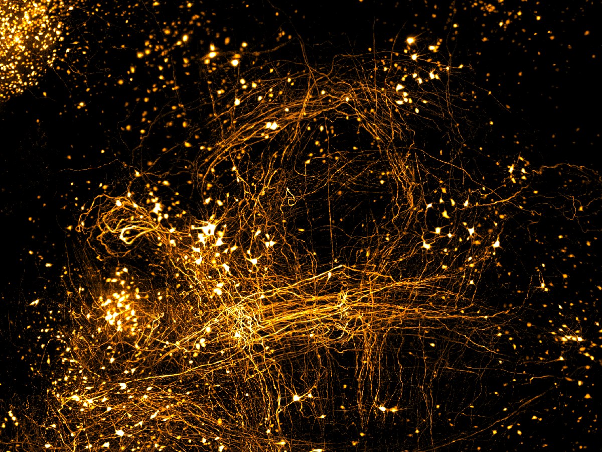
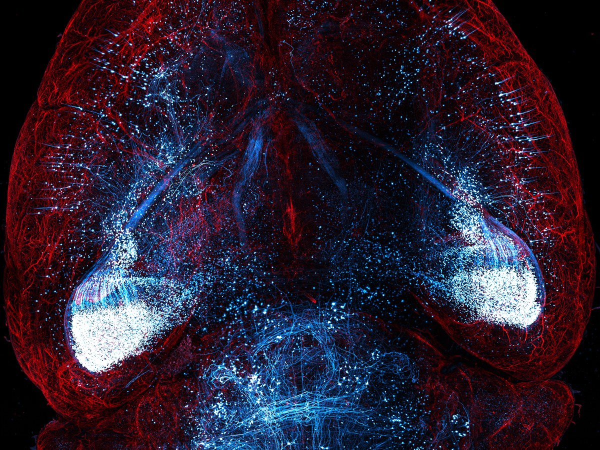
Intravital two-photon microscopy through thinned-skull cranial windows captures blood vessels and microglia as dynamic events, making cellular responses visible in the tissue environment where disease processes emerge.
Blood vessels are labeled red and microglia green during intravital imaging after noxious intranasal exposure.
Time-lapse microscopy reveals how vascular and immune-cell behavior can shift across minutes in a living cortical field.
Dynamic imaging captures brief events that are difficult to infer from fixed tissue alone.
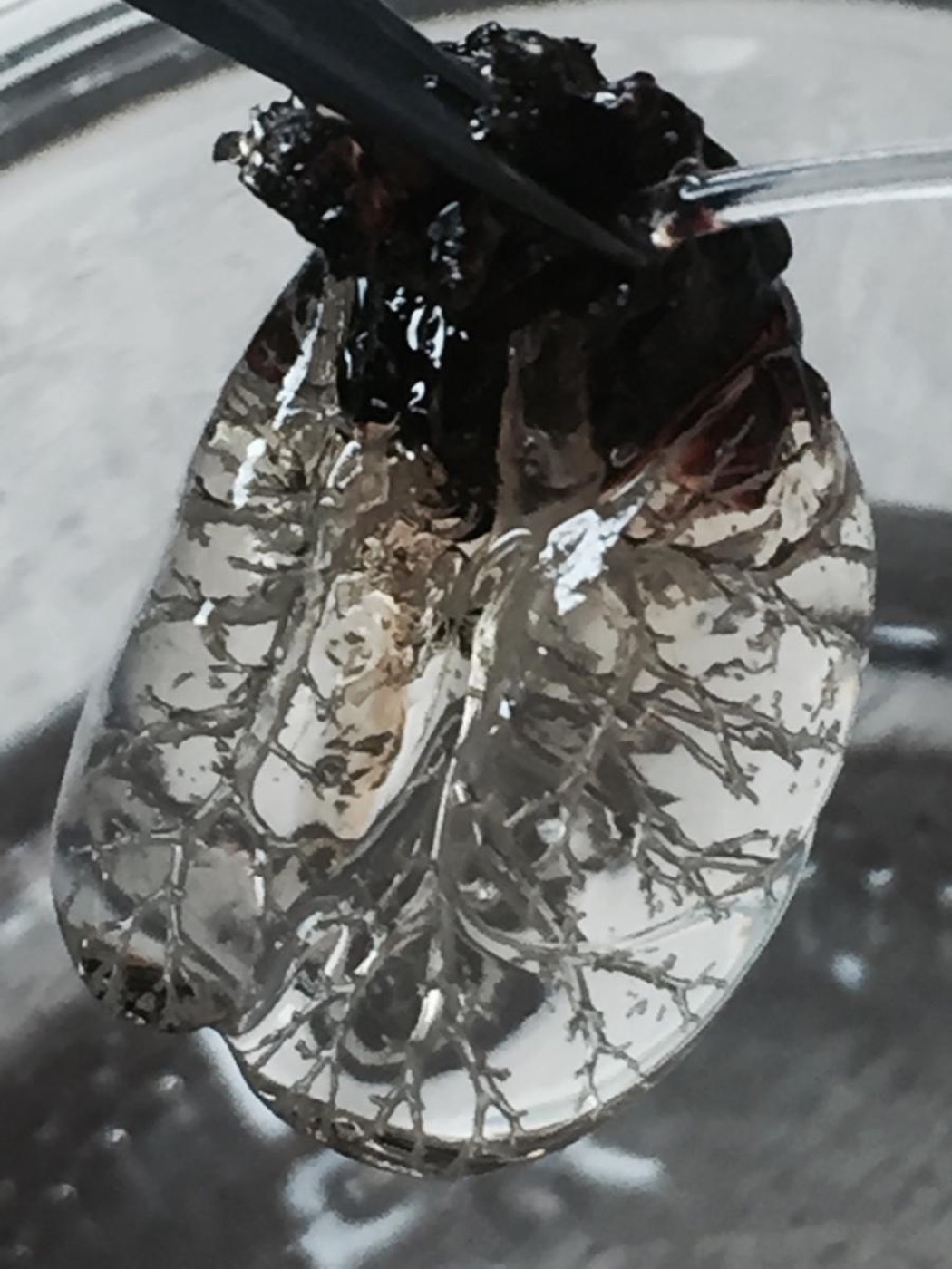
The lab's methodological foundation extends beyond thin-section imaging to intact organs, living tissue dynamics, and cleared specimens that preserve spatial relationships across scales.
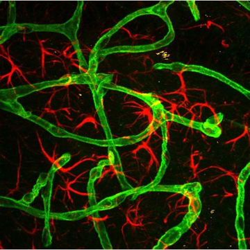
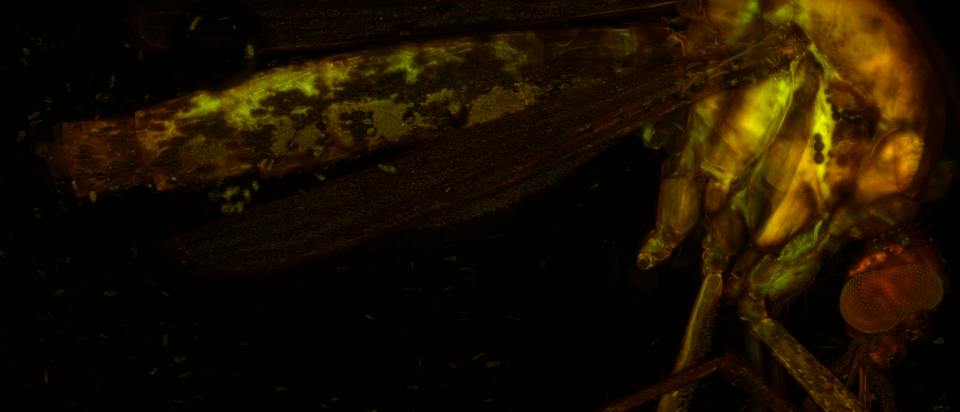
The lab's scientific questions depend on methods that preserve anatomical context while enabling rigorous comparison across scales, samples, and disease states.
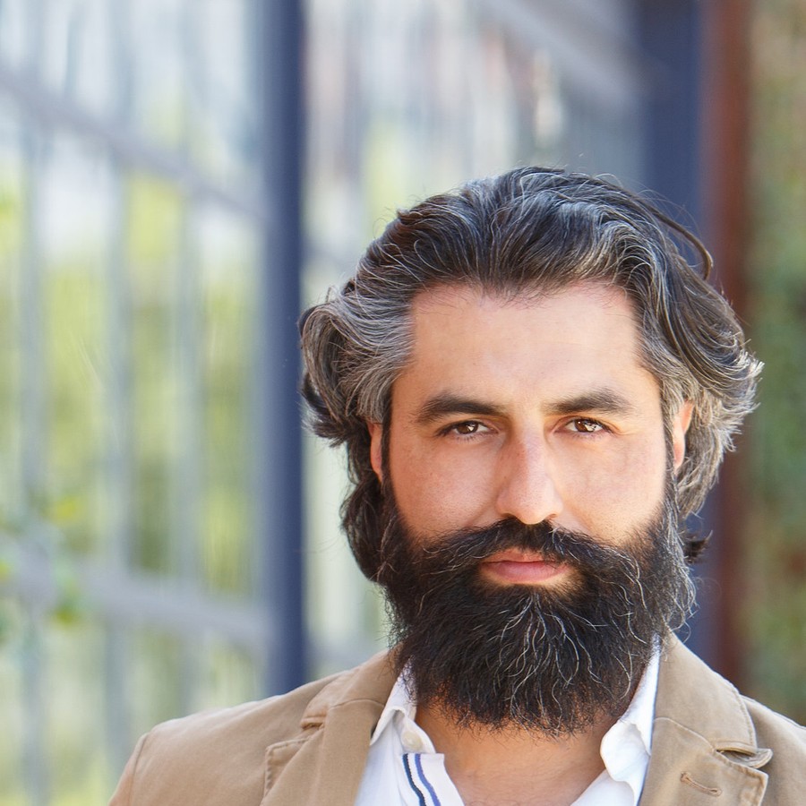
Lorenzo is a neuroscientist and bioengineer developing an independent research program at the intersection of circuit vulnerability, tissue transformation, spatial biology, and computational 3D pathology.
He is a Postdoctoral Fellow at MIT's Picower Institute for Learning and Memory in the Chung Lab, where he investigates systems-level mechanisms of neurodegenerative disease and the role of locus coeruleus circuitry in Alzheimer's pathology.
His multidisciplinary training spans neuroscience, biochemistry, biomedical engineering, optical clearing, whole-organ imaging, light-sheet and multiphoton microscopy, quantitative computation, infectious disease, and neuroinflammation.
Selected peer-reviewed work is shown below. Complete and current records are available through PubMed and Google Scholar.
Science is strongest when people feel intellectually challenged, professionally supported, and respected as whole individuals. The Ochoa Lab will aim to build a culture where rigorous research, technical creativity, openness, and care for one another are inseparable parts of the scientific process.
Regular one-on-one meetings, constructive feedback, project planning, and support for short-term milestones and long-term career goals.
Clear conversations about project goals, authorship, training needs, conference plans, fellowship applications, and progress.
High standards paired with documentation, reproducibility, clear communication, mutual respect, and lives outside the lab.
The lab is in development. Prospective trainees, research staff, computational collaborators, and scientific partners interested in neurodegeneration, neuroinflammation, volumetric imaging, tissue transformation, computational pathology, or spatial biology are welcome to reach out.
Contact usFormal positions and institutional details will be added once available.